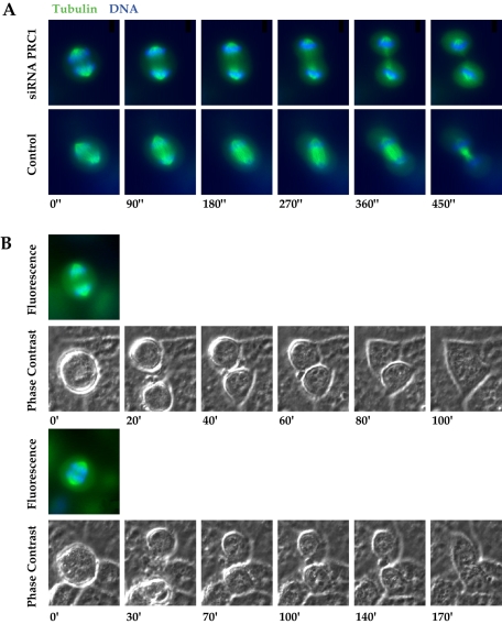
Figure 1.
Cells lacking a central spindle bundle undergo complete furrowing and then rejoin after failure of abscission. (A) Time frames from time-lapse videomicroscopy show progression of the cleavage furrow in a PRC1-ablated cell (top images) compared with a control (bottom images). Furrowing is complete in the absence of a central spindle bundle, and furrow progression occurs in approximately the same time course as in the control. Times indicated are in seconds. Images were collected of cells expressing EGFP-α-tubulin (green) and counterstained with Hoechst DNA dye (blue). The absence of a central spindle bundle is evident in the PRC1-ablated cell. See Video 1 for the full movie. (B) Cells that lack the central spindle bundle rejoin after furrowing is complete. Time frames from time-lapse videomicroscopy show phase contrast images of two cells that have undergone complete furrowing in the absence of a central spindle bundle and then rejoin more than an hour after furrowing has completed (time course to rejoining was variable but never <1 h after furrowing). Times indicated are in minutes. Color images above each initial frame show the same cells imaged for EGFP-α-tubulin (green) and counterstained with Hoechst DNA dye (blue) to indicate the absence of a central spindle bundle. The two cells were recorded in the same microscopic field. See Video 2 for the full movie.

