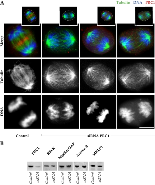
Figure 2.
Loss of the central spindle bundle but retention of anaphase spindle proteins after ablation of PRC1. (A) One control HeLa cell, and three examples of cells negative for PRC1 after siRNA ablation, are shown in anaphase (projections of 0.5-μm optical sections). In the merge, the control shows an accumulation of PRC1 (red) in the midzone of the mitotic spindle (green) and the segregated chromosomes (blue). In the absence of PRC1 (absence of red), chromosomes are segregated, although bridges are not established between the microtubules of the two half-spindles. Channel separations are shown to indicate in detail microtubule arrays in anaphase in the absence of PRC1. Insets above indicate the cell outlines in white. The siRNA cells are in late anaphase, whereas the control is in early telophase. Bar, 10 μm. (B) Western blots of HeLa cell extracts collected 24 h after transfection with PRC1 siRNA, compared with control HeLa at equivalent protein loadings. The PRC1 level is substantially suppressed by siRNA treatment at 24 h, whereas other proteins of the central spindle (MKLP1 and MgcRacGAP), and proteins required for cell cleavage (RB6K and Aurora B), are not diminished by ablation of PRC1.

