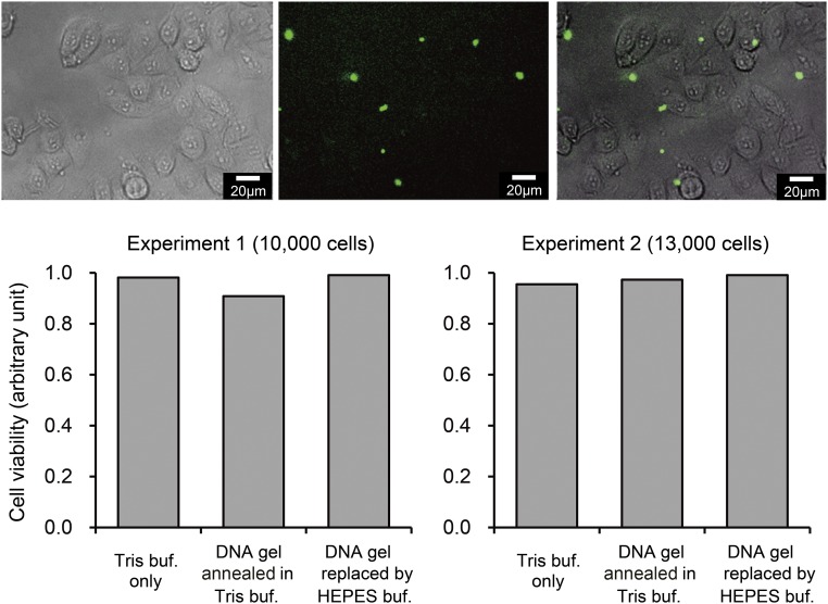
Fig. S4.
(Upper) Fluorescent and phase contrast microscopic images of cells after 24 h of incubation with DNA microgels (shown in green: annealing in Tris-buffer, then replaced by Hepes-buffer). (Scale bar, 20 μm.) (Lower) Cell viability after 24 h of incubation with DNA microgels, determined by the MTT assay (two independent experiments). Presence of DNA microgels did not significantly affect the viability of HeLa cells. buf, buffer.

