Figure 5.
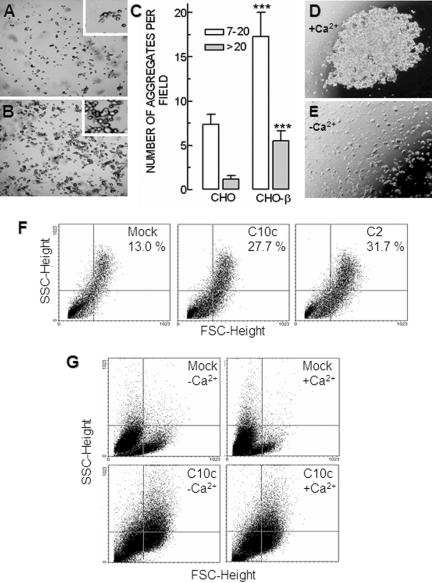
Cell aggregation assay. The adhesiveness of CHO cells transfected with the β-subunit was assayed following the two different aggregation assays described in Materials and Methods. (A) CHO cells transfected with the empty vector (mock) are mostly isolated or in small aggregates (inset). (B) CHO-β cells tend to aggregate in larger clumps (inset). (C) Clumps were scored into two groups: those with 7–20 cells (white bars) and those larger than 20 cells (gray bars). Groups of less than seven cells are not represented. The first two columns correspond to mock-transfected CHO cells that tend to group in small clumps of 7–20 cells. Transfection of β-subunit (last two columns) produces a 136% increase in the number of small clumps (p < 0.001) and a 380% in the number of large clumps (p < 0.001). Each column represents an average of 36 individual fields. MDCK cells at the tip of the hanging drop form a large clump in the presence of 1.8 mM of Ca2+ (D), whereas in the absence of Ca2+ the cells are dispersed (E). (F) Dot plots depicting cell complexity (SSC height) versus cell size (FSC height) of mock- and dog β1-transfected CHO cells (clones C10c and C2). All β1-transfected CHO cells exhibited at least a 100% increase in adhesiveness as estimated by the percentage of particles in the top right quadrant of the plot (higher size and complexity). (G) Dot plots of dog β1-(C10c) and mock-transfected CHO cells in the absence (-Ca2+) or presence (+Ca2+) of calcium ions in the bathing medium during aggregation assay.

