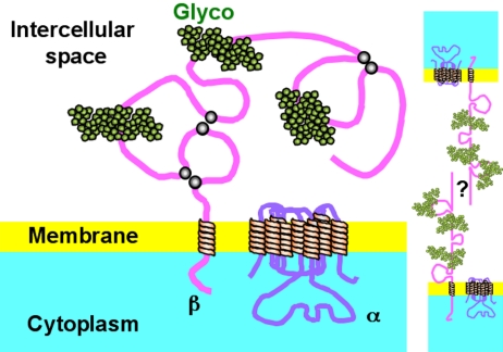
Figure 6.
Schematic representation of Na+,K+-ATPases located at the lateral border of epithelial cells. The α-subunit with its 10 transmembrane domains is represented. The β-subunit is depicted with a single transmembrane segment and a long extracellular domain containing three S-S links (gray dots) and three glycosylation sites (green). The structure and function of the enzyme requires that both subunits interact closely and strongly; yet for clarity, these are represented as molecules placed far from each other. For the same reason, γ-subunit is omitted. On the right-hand side two β-subunits belonging to neighboring cells are represented as spanning the intercellular space. The present results suggest the possibility that a β-β linkage anchors the Na+,K+-ATPase at the lateral borders of epithelial cells. The question mark indicates that we ignore whether the β–β interaction would be a direct one or mediated by an as yet unknown molecule.

