Fig. 6.
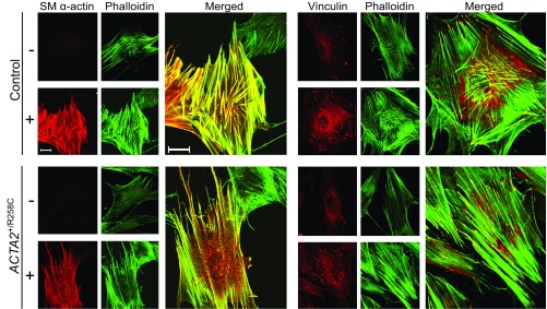
Induction of SM α-actin in R258C cells results in less prominent stress fibers and focal adhesions than in control cells. Representative confocal micrographs of control (Upper) and R258C (Lower) cells infected with (+) or without (−) Ad-MRTF-A for 48 h followed by subculture overnight with fixation and staining as described. (Left set) SM α-actin immunofluorescence (red) and Alexa-488 phalloidin-stained F-actin (green). Enlarged image represents merge of MRTF-A−–induced cells. (Right set) Representative confocal micrographs of vinculin immunofluorescence (red) and Alexa-488 phalloidin-stained F-actin (green). Enlarged image represents merge of MRTF-A–induced cells. (Scale bars, 20 μm.)

