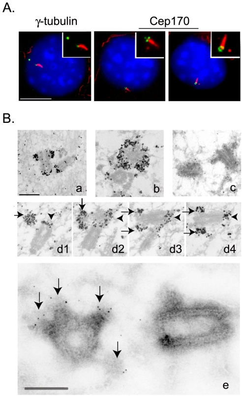
Figure 7.
Localization of Cep170 to appendages on the mature centriole. (A) Costaining of serum-starved NIH 3T3 cells with antibodies against acetylated tubulin (red) and either γ-tubulin or Cep170 (green), as indicated. Insets show enlargements of centrosomes and cilia. Bar, 10 μm. (B) Immuno-EM localization of Cep170 in U2OS cells. Preembedding (a–d): longitudinal section of one centrosome (a); transversal section of one centriole with the typical “crown” labeling produced by the anti-Cep170 antibody (b); control staining with secondary antibody only (c); and serial longitudinal sections through one centrosome (arrows indicate labeling of appendages, arrowheads mark the proximal staining at both centrioles) (d1–d4). Ultrathin cryosection (e), enlarged image of one centrosome; arrows indicate labeling of appendages. Bars, 250 nm.

