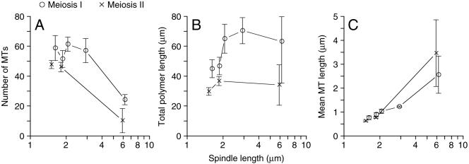
Figure 3.
General description of the microtubules in MI (○) and MII (×) spindles derived from the 35 meiotic spindles (see Table 1) as described in Materials and Methods. (A) The total number of microtubules versus spindle length. (B) The microtubule total polymer versus spindle length. (C) Mean microtubule length versus spindle length. Spindles of similar length were grouped together. Error bars represent 74% confidence interval.

