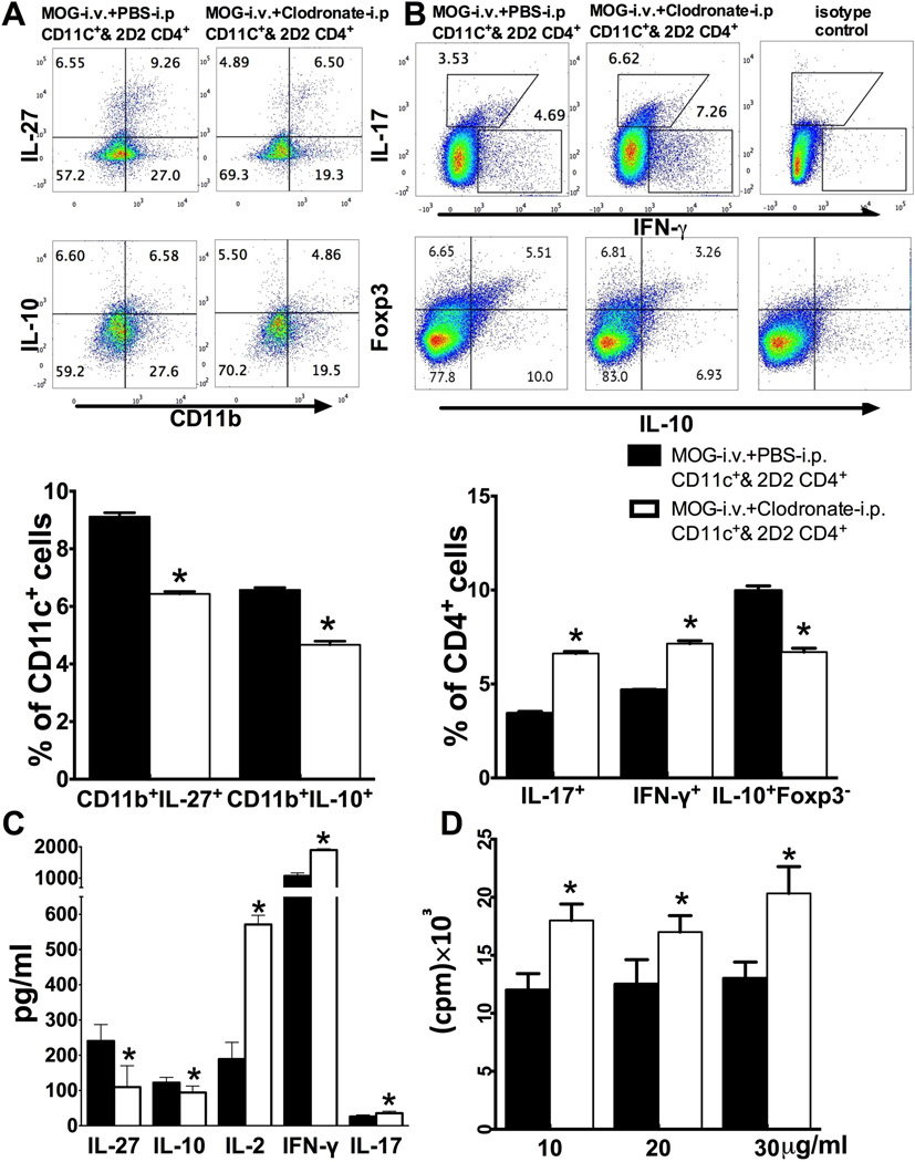
Fig. 7. Effect of iDC depletion on MOG-reactive T cells in co-culture.
CD4+ T cells from naïve spleens of MOG TCR transgenic mice (2D2) were co-cultured with DCs from spleen of EAE mice treated with MOG-i.v.+PBS-i.p. (MOG-i.v.+PBS-i.p. DCs & 2D2 CD4) or MOG-i.v.+clodronate-i.p. (MOG-i.v.+clodronate-i.p. DCs & 2D2 CD4) in the presence of MOG35–55 for 3 days. (A) CD11c+ cells were gated and the intracellular secretion of IL-27 and IL-10 in CD11c+CD11b+ cells were analyzed (up) and expressed as mean ± SEM (n=3 each group; bottom). (B) CD4+ cells were gated and their intracellular IFN-γ, IL-17, Foxp3 and IL-10 were analyzed with flow cytometry (top) and data are shown as mean ± SEM (n=3 each group; bottom). (C) Culture supernatants were harvested and production of IL-27, IL-10, IL-2, IFN-γ, IL-17 was analyzed with ELISA. Data are expressed as mean ± SEM (n=4 each group). (D) MOG-induced T-cell proliferative responses were measured by 3H-methylthymidine incorporation and the values of cpm (counts per minute) are shown as mean ±SEM (n=3 each group). *p<0.05, unpaired Student’s t test. One representative of 3 experiments is shown.

