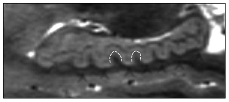
Figure 1.

This image depicts an ultra-high resolution sagittal view of the hippocampus, which is necessary to clearly differentiate the gray matter of CA1/subiculum and CA4/hilar region from the dark band of white matter constituted by the strata radiatum, lacunosum, and moleculare (SRLM). Arrows indicate the dark SRLM layer and the CA1/subiculum and CA4 areas; arrowheads indicate the dentes on the hippocampal body and tail, of interest in this study.
