
Figure 3.
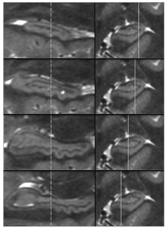
Panels on the left show sagittal views of the hippocampus at locations from lateral to medial as indicated by the solid lines in the panels on the right. The dashed lines indicate the coronal plane of section of the images on the right, which is identical in A-D. A single dente indicated by the arrows shows that it is visible in the lateral-most pane (A) and through the inferior aspect of the hippocampus (B & C), but is not apparent in pane D at this point along the anterior-posterior axis of the hippocampus. However, in D more posterior dentes can be seen because dentation shifts from being most prominent in the infero-lateral aspect anteriorly to the infero-medial aspect posteriorly.