
Figure 4.
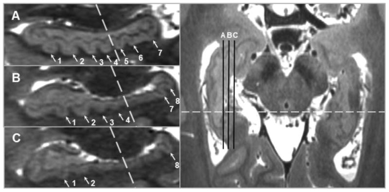
Dentation is assessed by scrolling through sequential sagittal slices because specific dentes may be only visible in more lateral or more medial slices, particularly in the hippocampal tail. The position of sagittal slices A, B, and C are indicated by the black lines in the axial slice in the right panel; the dashed lines indicate the boundary between anterior and posterior. Slice A shows 4 anterior and 3 posterior dentes (1-7), while in Slice B the first two posterior dentes cannot be seen (5-6) but an eighth emerges, and in Slice C only the eighth posterior dente is visible.