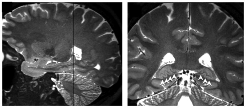
Figure 5.

Image A depicts a sagittal view of prominent posterior hippocampal dentation. B shows the corresponding coronal view (location indicated by the dashed line in A). This illustrates how posterior hippocampal dentation is often viewed more clearly in the coronal plane due to the inward (medial) curvature of the hippocampal tail. Dentation is shown with white arrowheads in B.
