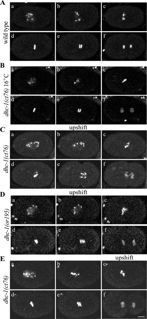
Figure 6.
Defects in chromosome congression in dhc-1 embryos upshifted during prometaphase. Time-lapse confocal images show embryos expressing GFP::histone. (A and Video 10) In wild-type embryos incubated at either 16 or 25°C, chromosomes congressed to form a tight metaphase plate (d) and segregated to form two discrete and well ordered groups (e and f). (B) In dhc-1(ct76) mutant embryos maintained at 16°C, chromosome congression (c and d) and segregation (e and f) looked normal. (C and Video 11) In dhc-1(ct76) mutant embryos shifted from 16 to 25°C at NEB, chromosomes congressed poorly (c and d), chromosomes lagged during anaphase (e), and segregation was defective (e and f). (D and Video 12) In dhc-1(or195) mutant embryos shifted from 16 to 25°C at NEB, chromosome congression was defective (c and d) and some chromosomes lagged in early anaphase (e), but segregation was better than in ct76 embryos (f). (E and Video 13) In dhc-1(ct76) embryos shifted from 16 to 25°C in late prometaphase during congression, chromosomes did not fully congress to a tight metaphase plate (d). Some lagging chromosomes were observed in early anaphase (e), but the subsequent extent of separation and cytokinesis seemed normal (f). Bar, 10 μm.

