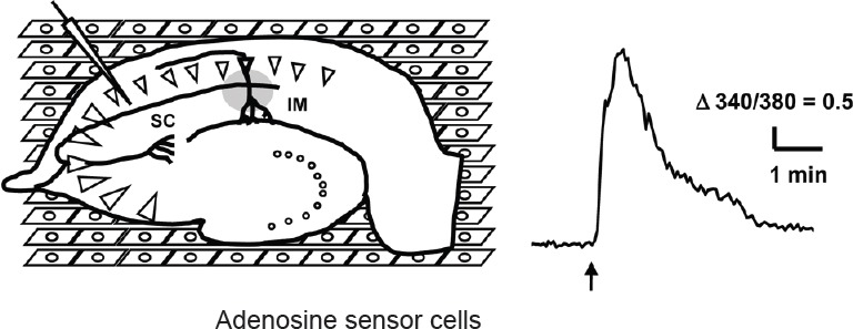
Figure 1.

Measurement of adenosine release in hippocampal slice following electrical stimulation by adenosine sensor cells.
A hippocampal slice was placed on the top of the adenosine sensor cells (human embryonic kidney 293 (HEK293) cells expressing A1 receptor and Gqi5) loaded with Fura-2AM and a high-frequency electrical stimulation was delivered to schaffer collateral (SC) (left). Calcium response of an adenosine sensor cell imaged by an inverted microscope (IM) following electrical stimulation (arrow) (right).
