
Figure 2.
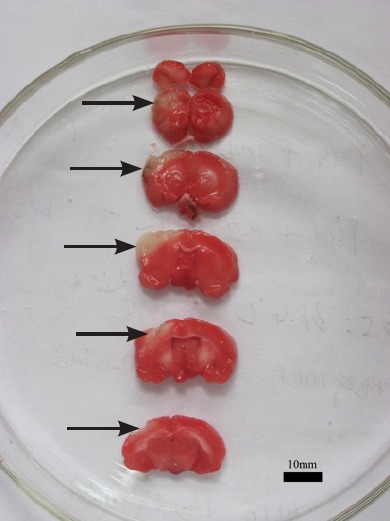
Pathological change of cerebral ischemia/reperfusion rats (2,3,5-triphenyltetrazolium chloride staining).
The white parts of the brain tissue are the infarcts, and their locations are in the area of middle cerebral artery.
Official websites use .gov
A
.gov website belongs to an official
government organization in the United States.
Secure .gov websites use HTTPS
A lock (
) or https:// means you've safely
connected to the .gov website. Share sensitive
information only on official, secure websites.
Pathological change of cerebral ischemia/reperfusion rats (2,3,5-triphenyltetrazolium chloride staining).
The white parts of the brain tissue are the infarcts, and their locations are in the area of middle cerebral artery.