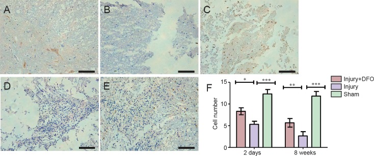
Figure 5.
DFO effects on the number of NeuN(+) neurons in injured spinal cord of rats.
(A–E) Images of immunohistochemistry sections obtained using an inverted fluorescence microscope. Scale bars: 100 μm. (A) Normal neurons are observed 2 days post injury in the sham group. (B) By contrast, the neurons appeared to have shrunken, the tissue structure is damaged, and there is inflammatory cell infiltration 2 days post injury in the injury group. (C) Residual neurons are observed in the injury + DFO group 2 days post injury. (D) However, there are fewer neurons in the injury group compared with injury + DFO group 8 weeks post injury. (E) In the injury + DFO group 8 weeks post injury, there is an evident restoration of neuronal number and morphology compared with injury group. (F) The number of NeuN-positive cells was counted in the three groups and found to be significantly different between the injured saline- and DFO-treated rats at both 2 days and 8 weeks post injury (*P < 0.05, **P < 0.01, ***P < 0.01 for the comparisons indicated). Data are expressed as the mean ± SEM (n = 3 per group, one-way analysis of variance followed by Tukey's post hoc test). DFO: Deferoxamine.

