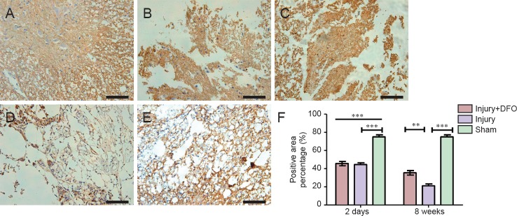
Figure 6.
DFO effects on the number of oligodendrocytes in injured spinal cord of rats (inverted fluorescence microscope).
(A–E) Spinal cord samples collected 2 days (A, B, C) and 8 weeks (D, E) post injury. Scale bars: 100 μm. The CNPase(+) area shows normal morphology in the sham group (A). The tissue structure is damaged, the percentage of the CNPase(+) area is lower compared with the sham group, and the CNPase(+) area is atrophic in the injury group (B). The CNPase(+) area in the sham group has a normal structure, while in the injury group (D), the tissue structure is damaged, with a lower percentage of the area that is CNPase(+). The percentage of the CNPase(+) area and the tissue structure in the CNPase(+) area of the injury + DFO group (E) is larger than that in injury group. (F) There are no differences in the percentages of CNPase(+) areas between the injured saline- and DFO-treated rats at 2 days, but these groups are significantly different at 8 weeks post injury (**P < 0.01, ***P < 0.001 for the comparisons indicated; mean ± SEM, n = 3 per group, one-way analysis of variance followed by Tukey's post hoc test). DFO: Deferoxamine.

