Figure 2.
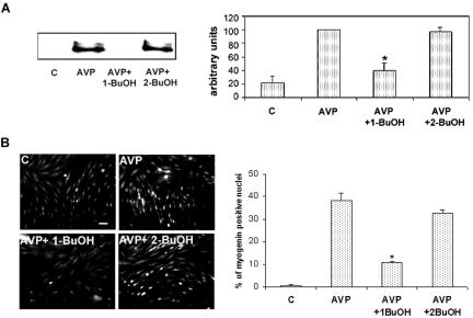
AVP induces myogenin expression and nuclear accumulation in a PLD-dependent way. (A) Immunoblots of myogenin from L6 cells treated for 48 h by AVP alone or in the presence of 0.5% 1-butanol or 0.5% 2-butanol. Videodensitometric quantitation of myogenin protein: the blots were reprobed for tubulin, and myogenin amounts were normalized by tubulin. The average of three different experiments is shown in the diagram. *, different from AVP alone, p < 0.01. (B) Immunofluorescence of myogenin in L6 cells treated for 48 h by AVP alone, or in the presence of 0.5% 1-butanol or 0.5% 2-butanol. Nuclear myogenin was revealed by using a monoclonal antimyogenin antibody and a fluorescein-conjugated secondary antibody. The total number of nuclei was evaluated on the phase contrast image. The average percentages of myogenin-positive nuclei counted in five to nine different fields (∼70 cells per field) in one experiment are shown in the diagram. *, different from AVP alone, p < 0.01. Three experiments gave similar results. Full reversibility of the effects of a 15-min treatment by 1-butanol on myogenin nuclear accumulation was verified (our unpublished data). Bar, 40 μm.

