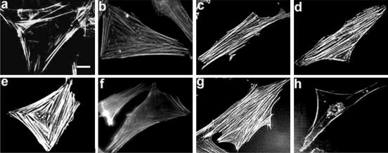
Figure 6.
Stress fiber-like actin structures are formed in a PLD-dependent way in L6 cells stimulated by AVP. Cells treated for 0–60 min by AVP, were fixed, labeled with rhodamine-phalloidin, and observed by fluorescence microscopy (a–e, times 0, 30 s, 1 min, 10 min, and 60 min). The effects of a cotreatment by 1% 1-butanol (f) or 1% 2-butanol (g), or of a pretreatment by C3 exoenzyme (h) on the SFLS formation induced by a 10-min AVP treatment are shown. SFLSs were present in 56 ± 6% of AVP-treated cells after 10 min versus 2.0 ± 0.9% of control cells. In the presence of 1-butanol, only 1.6 ± 0.6% of AVP-treated cells displayed SFLSs versus 40 ± 3.3% for 2-butanol + AVP-treated cells (means ± SE of 10 fields, ∼300 cells). The inhibition of SFLS formation by butanol was reversed by washing out the alcohol (our unpublished data). Bar, 5 μm.

