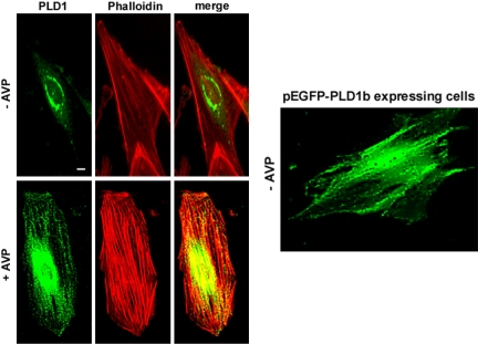
Figure 7.
Localization of endogenous and overexpressed PLD1 in L6 cells. Quiescent (top left) or 10 min AVP-stimulated cells (bottom left) were fixed, and PLD1 was detected by immunofluorescence by using a specific antibody. Colabeling of actin was performed with phalloidin, and the images were merged. Fluorescence of GFP-PLD1b expressed in live cells stimulated for 10 min by AVP was examined (right). Bar, 5 μm.

