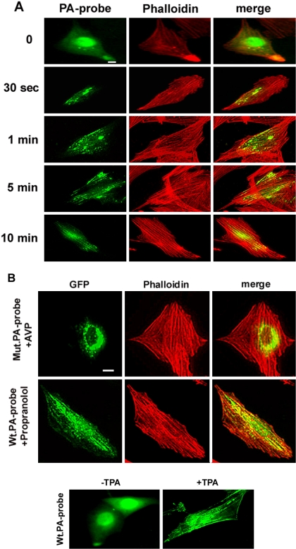
Figure 8.
Labeling of PA with an endogenously expressed PA-specific fluorescent probe. (A) L6 cells expressing the Raf1 PA-binding domain fused to GFP (PA-probe) were examined for green fluorescence and for rhodamine-phalloidin–stained actin, after treatment by AVP for the time indicated. The images were merged. (B) A probe mutated for arginine residues essential for PA binding (mutPA-probe) was expressed in the cells. After a 10-min AVP stimulation, the cells were examined as described above (top). L6 cells expressing the unmutated PA-probe were treated by 100 μM propranolol for 15 min, before being examined as above (middle). In the presence of propranolol, 24 ± 2.0% of the cells expressed SFLSs versus 3.5 ± 1.1% of untreated cells (means ± SE of 10 fields, ∼200 cells). L6 cells expressing the unmutated PA-probe were treated or not (control) by 1 nM TPA for 15 min, before being examined for probe fluorescence (bottom). Bar, 5 μm.

