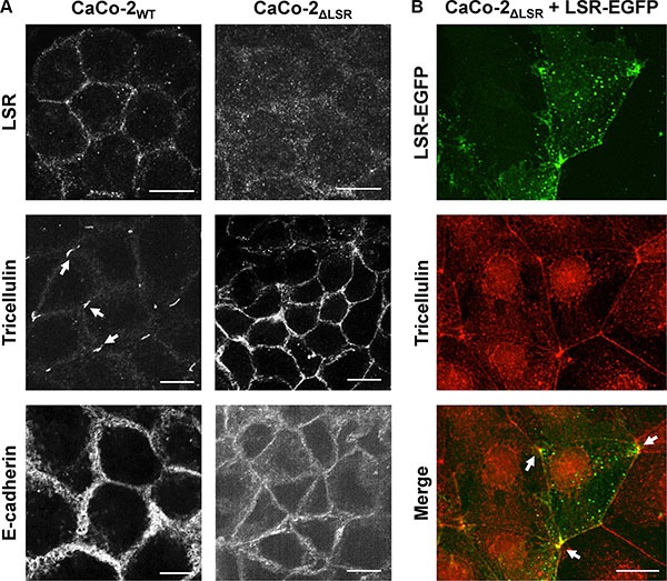
Figure 7. Staining of bi- and tricellular marker proteins in cell monolayer of wild-type and LSR-deficient CaCo-2 cells.

(A) Immunostaining of LSR (top panel), tricellulin (middle panel) and E-cadherin (bottom panel) in CaCo-2WT and CaCo-2ΔLSR cells with specific antibodies and analysis of the intracellular distribution of the respective proteins via confocal fluorescence microscopy. (B) Reconstitution of LSR expression in CaCo-2ΔLSR cells. CaCo-2ΔLSR cells were transfected with a plasmid that leads to ectopic expression of LSR-EGFP (green signal). Cells were immunostained for tricellulin (red signal). Arrows indicate accumulation of LSR-EGFP and tricellulin in tricellular contacts. Scale bars represent 10 μM.
