
Figure 4. Combination treatment with NK-92MIhCD16 cells and rituximab successfully suppressed the growth of a CD20-positive tumor in a xenograft mouse model.
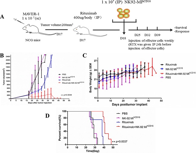
(A) Experiment schema: Seven-week-old female NCG mice were injected subcutaneously in the dorsa; right side with 1 × 10⁷ MAVER-1 cells. When tumors reached a volume of 200 mm3, mice were individually identified and randomly assigned to the control or treated groups (4–6 mice per group) and treatments were initiated (all mice were sacrificed before the tumor volume reached 2500 mm3). Rituximab (400 μg/mouse) was injected intraperitoneally once weekly for four weeks starting on day 17. Statistical analysis was performed using the log-rank test. (B) Tumor volume of the mice was measured every 2–3 days (**P < 0.01). (C) Body weights of the mice were measured every 2-3 days. The values are presented as the mean ± standard error of the mean. (D) Kaplan–Meier survival curve for mice treated with NK-92MIhCD16 cells in combination with rituximab compared to control treated mice. NK-92MIhCD16 cells in combination with rituximab treated mice survived significantly longer than the control. Log-rank (Mantel-Cox) test was used to determine significance. (**P < 0.01).