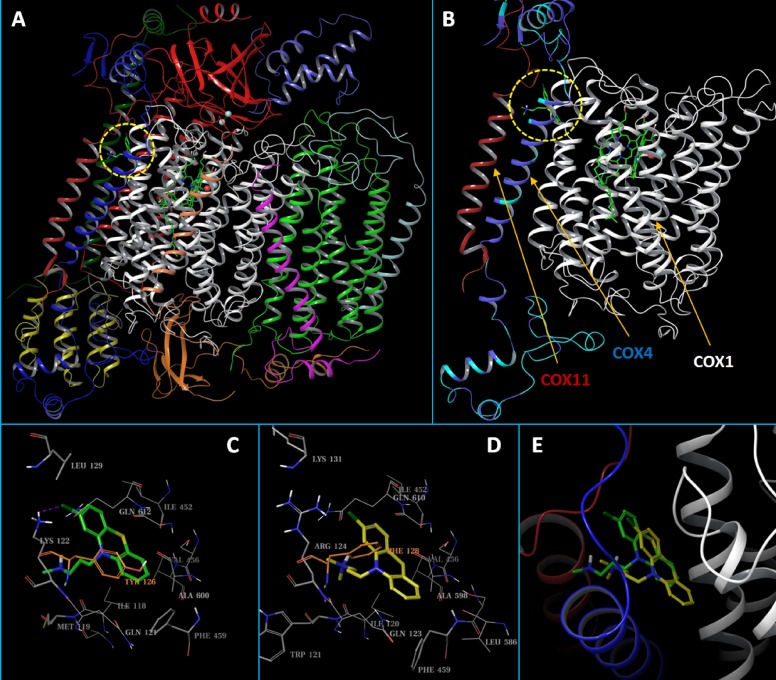
Figure 8. Structural presentation of the predicted binding modes of CPZ to human CcO.
(A) Carton presentation of the CcO complex model with each of its 13 subunits shown in differently colored ribbons. The predicted binding site, siteA, was circled in a dashed yellow line. (B) CcO subunits that are close to siteA. The HEME molecule and the docked CPZ molecule are shown in solid sticks. COX1 and COX11 are shown in white and red ribbons, respectively. For the COX4 ribbon, residues that are the same between COX4-1 and COX4-2 were colored in blue, while residues that are different are colored in cyan. (C and D) Close up view of the binding site interactions of the docked CPZ in the COX4-1 and the COX4-2 models, respectively. The CPZ molecules are shown in solid sticks and colored in green and yellow, respectively. Binding site residues are shown in gray lines. The key and non-conserved residues are shown in thin sticks, or colored in orange. (E) Overlaid docked CPZ molecules from the COX4-1 and COX4-2 models. CPZs and CcO ribbons are colored the same as above.

