
Figure 3. NK cell function was impaired via exosome.
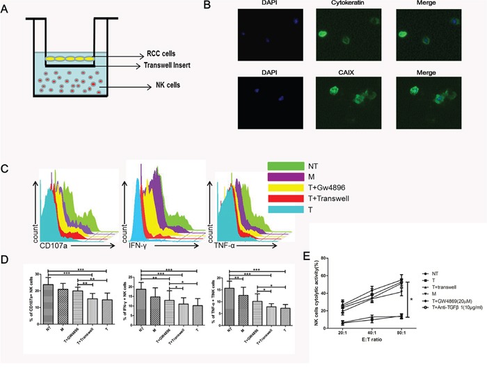
(A) Transwell coculture model was used for the co-culture of primary NK cells and ccRCC cells. (B) Representative micrographs of immunofluorescence staining of cytokeratin and CAIX. DAPI counterstains nuclei in blue. 200× magnification. (C, D) NK cell functions (CD107a degranulation, IFN-γ and TNF-α production) in NT, M, T+GW4869, T+transwell and T groups. (E) Cytotoxicity of NK cells coculture with NT, M, T+GW4869, T+transwell, T and anti-TGFβ1 groups. All data shown were from three independent experiments. Error bars indicate ±SD, *p < 0.05, **p < 0.01 and ***p < 0.001vs corresponding control.