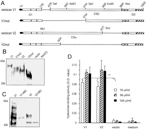
Figure 1.
Generation and expression of versican constructs. (A) Full-length versican V1 and V2 isoforms and two mutants (V1mut and V2mut; derived from V1 and V2 isoforms, respectively) were generated as shown in the figure. (B) Stable expression of V1, V2, V1mut, and V2mut in NIH3T3 fibroblasts was assessed by Western blot probed with the monoclonal antibody 4B6 that recognizes an epitope on the leading peptide (LP) of each construct. (C) The products of V1 and V2 were digested with chondroitinase ABC followed by Western blot analysis to confirm the removal of GAG chains from the product. (D) To test the binding activity of the secreted proteoglycan, hyaluronan, which binds the G1 domain of versican, was coated on ELISA plates at different concentrations followed by incubation with fresh medium or culture medium collected from cells transfected with V1, V2, or the control vector. Bound products were detected with a polyclonal antibody that recognizes the G3 domain of versican. The V1 and V2 products exhibited hyaluronan-binding activity (n = 3, **p < 0.01).

