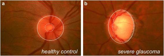
Figure 4.

Fundoscopic image from a healthy patient with a small cup-to-disc ratio and a healthy, pink optic disc surrounding the cup (a) compared with a fundoscopic image from a severely glaucomatous patient with an enlarged cup and significant inferior thinning of the disc rim (b). Dashed white circles roughly outline the cups; solid white circles roughly outline the discs.
