Figure 4.
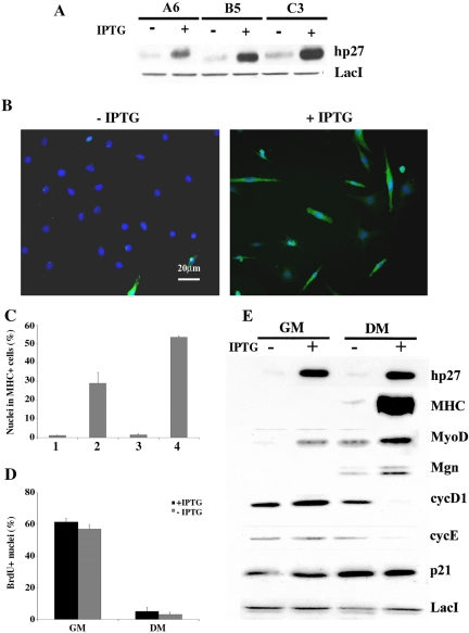
Forced expression of p27Kip1 recovers the ability of C2C12 myoblasts to differentiate at low density culture conditions. (A) Exogenous p27Kip1 expression in three engineered clones determined after 24 h in GM minus (-) or plus (+) IPTG. (B) A6 cells, plated at LD, were induced to differentiate in the absence or the presence of IPTG and MHC expression was analyzed after 5 d by immunofluorescence (green). Nuclei were counterstained in blue (Dapi) and individual pictures of the same field, taken with a DC camera, were merged using a LEICA Microsystems Imaging Equipment. (C) Analysis of MHC positive A6 cells following addition of IPTG at different times: for only 24 h in GM (column2),for 5 d in DM (column 3), or throughout the whole experiment, 24 h in GM + 5 d in DM, (column 4). Column 1 refers to untreated cells. (D) The percentage of cells in S phase was determined after 2 h BrdU labeling (50 μM) by immunofluorescence staining. (E) Protein extracts from A6 cells grown in GM for 24 h minus or plus IPTG or in DM for 5 d minus or plus IPTG were probed with the indicated antibodies. The expression of LacI repressor was used to normalize the amount of protein loaded. Data are the means ± SD of three independent experiments.

