Figure 7.
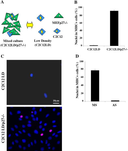
Cell-cell contact with fibroblasts is sufficient to induce terminal differentiation of C2C12 cells cultured at LD. (A) Schematic representation of the experimental setting: C2C12A5LacI (simply named C2C12) cells cultured alone at LD (C2C12LD) or in mixed culture with an excess of mouse embryo fibroblasts from p27Kip1 knock out mice (C2C12LD/p27-/-). (B) C2C12 cells seeded as shown in (A) were shifted to DM 24 h after plating. 3 d later the incidence of differentiated C2C12 cells were evaluated by MHC staining. Data are the means ± SD of three independent experiments. (C) p27Kip1 expression in C2C12 myoblasts cultured at LD alone or in mixed culture with MEFp27-/- after 24 h in GM. Nuclei were counterstained in blue (Dapi); the pink colored nuclei result from the overlapping of the red (anti-p27Kip1) and blue (DAPI) colors and represent the double-stained cells. (D) C2C12LD/p27-/- mixed cultures were transfected in GM with p27Kip1 antisense (AS) or mismatch (MS) oligonucleotides and shifted to DM after 24 h. 3 d later the incidence of differentiated C2C12 cells was evaluated by MHC staining. Data are the means ± SD of three independent experiments.

