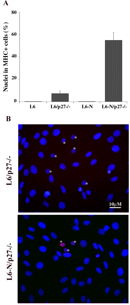
Figure 8.
N-cadherin-mediated cell-cell contact allows p27Kip1 accumulation and terminal differentiation in LD myoblasts. (A) L6C5 and L6C5-N rat myogenic cells were plated at LD alone (L6 and L6-N) or in mixed culture with an excess of MEFp27-/- (L6/p27-/- and L6-N/p27-/-) and, after 24 h in GM, were placed in DM for 4 d. The incidence of differentiated cells was evaluated by MHC staining. (B) The expression of p27Kip1 was detected after 24 h in GM by immunofluorescence with specific antibodies. Nuclei were counterstained in blue (Dapi). The pink colored nuclei result from the overlapping of the red (anti-p27Kip1) and blue (DAPI) colors. Rat nuclei (*) can be distinguished from mouse nuclei characterized by intensely fluorescent chromocenters.

