
Figure 12.
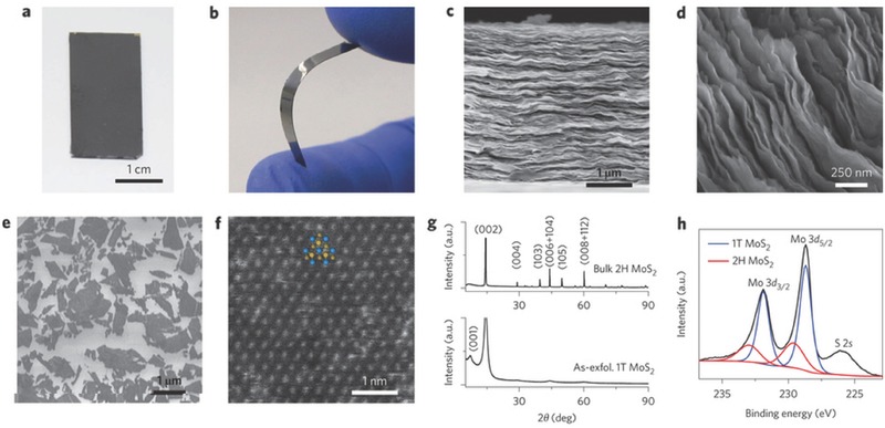
Chemically exfoliated 1T MoS2 electrodes. a,b) Photographs of electrodes consisting of a thick film of chemically exfoliated 1T MoS2 prepared by vacuum filtration and transferred onto rigid glass (a) and a flexible polyimide substrate (b). c) Side view of the electrode observed by scanning electron microscopy (SEM) showing the layered nature of the film made by restacking exfoliated MoS2 nanosheets. d) High‐magnification image of restacked MoS2 nanosheets. e) SEM image of as‐exfoliated monolayer 1T phase MoS2 nanosheets. f) High‐angle annular dark‐field scanning transmission electron microscope image of monolayer 1T phase MoS2. Inset: Atomic structure of 1T phase MoS2 (Mo and S atoms are displayed in blue and yellow, respectively). g) XRD of bulk MoS2 compared with as‐exfoliated restacked MoS2. h) High‐resolution X‐ray photoelectron spectrum from the Mo 3d region of as‐exfoliated 1T MoS2 (black). Contributions from 1T and 2H phase components in the Mo 3d spectrum are indicated by blue and red curves, respectively. Reproduced with permission.260 Copyright 2015, Nature Publishing Group.