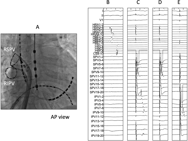
Figure 3.
Representation figure of EEPVI with interpulmonary isthmus ablation. (A) Catheter positions during right PV isolation. (B) Intracardiac electrogram of the right superior (RSPV) and inferior (RIPV) pulmonary veins just after EEPVI. No PV potential was observed in both PVs. (C) Pacing at the superior PV by using a ring catheter. A local electrogram of the inferior PV was captured. (D) After interpulmonary isthmus ablation, local electrogram of the inferior PV was not captured despite pacing at the superior PV. (E) Local electrogram of the superior PV was not captured despite pacing at the inferior PV, hence the bidirectional block of the interpulmonary isthmus. AP; anterior-posterior ,EEPVI; extensive encircling pulmonary vein isolation, PV; pulmonary vein.

