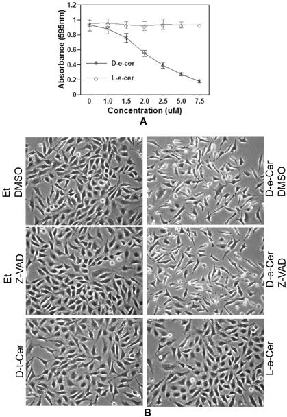
Figure 1.
(A) Ceramide treatment causes growth arrest and morphological changes of HeLa cells. HeLa cells grown to a 60% confluence in MEM medium with 10% FBS were treated with various concentrations of D-e-Cer or L-e-Cer for 24 h and were subjected to MTT assays for viable cell number. (B) HeLa cells treated with 2.5 μM D-e-Cer, 10 μM L-e-Cer, 10 μM D-t-Cer, or ethanol (Et) for 12 h in the presence or absence of 50 μM Z-VAD-fmk were imaged under an inverted microscope (Nikon Eclipse TE200) equipped with a CCD camera (Dage MTI 330T-RC). Data represent mean value ± SD of three different experiments performed in duplicate. This figure is representative of at least three independent experiments.

