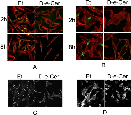
Figure 7.
Ceramide alters the arrangement of the microtubule and F-actin cytoskeletons and inhibits the formation of focal adhesions and filopodia. After being treated with 2.5 μM D-e-Cer or Et for different times, HeLa cells were stained with antibodies against α-tubulin (red) and giantin (green) (A), phalloidin-rhodamine (red) and giantin (green) (B), or paxillin (C) before confocal microscopic analyses. Note that the vehicle-treated cells contain abundant filopodia that are significantly reduced in the ceramide-treated cells. (D) After 24 h transfection with the plasmid pEYFP-Mem, which directs expression of enhanced yellow fluorescent protein (EYFP) targeting to the plasma membrane, HeLa cells were treated for D-e-Cer or vehicle control for 12 h before confocal microscopic analysis. Note that filopodia were significantly inhibited in the D-e-Cer–treated cells. This figure is representative of at least three independent experiments.

