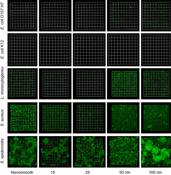
Figure 1.

Constructed confocal laser scanning microscopy (CLSM) three-dimensional images of 48-hour-old biofilms of E. coli O157:H7, E. coli K12, L. monocytogenes, S. aureus and S. epidermidis on nanosmooth alumina (control) and anodised surfaces of 15, 25, 50 and 100 nm pore diameter. The presented images have biomass accumulation close to the average for their surface type, so that images are representative. Scale units (small grid) are 34 μm in length.
