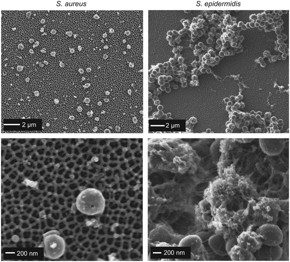
Figure 3.

Scanning electron microscopy images of S. aureus (left images) and S. epidermidis cells (right images) at low magnification (upper) and high magnification (lower) after 48 h contact time with anodic alumina surfaces with 100 nm pore diameter. Bottom right image shows S. epidermidis cells entrapped in a matrix of extracellular material.
