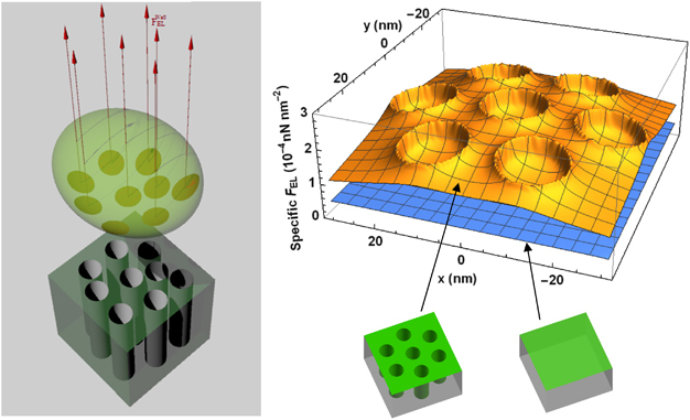
Figure 4.

Electrostatic repelling force field exerted on bacterial cells by the nanoporous surface. Left: schematic representation of the electrostatic repelling forces exerted on a bacterial cell by the nanopores located directly underneath the cell. Right: spatial distribution of the electrostatic repulsive force field exerted on E. coli O157:H7 cells by an anodic alumina surface with cylindrical pores of 15 nm diameter and 2,259 nm pore depth (top plot) compared with a smooth alumina surface (bottom plot), at a cell–surface separation distance of 0.2 nm. The surface components contributing to the force fields are illustrated in green.
