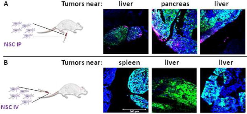
Figure 5. Neural stem cell administration route to peritoneal ovarian cancer metastasis.
NSCs labeled with CellMask Deep Red plasma membrane stain (magenta) demonstrate good distribution when (A) administered IP but not when (B) administered IV in a mouse model of peritoneal ovarian metastasis established using OVCAR8.eGFP.ffluc cells. (2 million NSC.CellMask in 200 µL PBS injected IP on Day 21; then harvested 4 days post-NSC injection). Scale bars = 500 microns.

