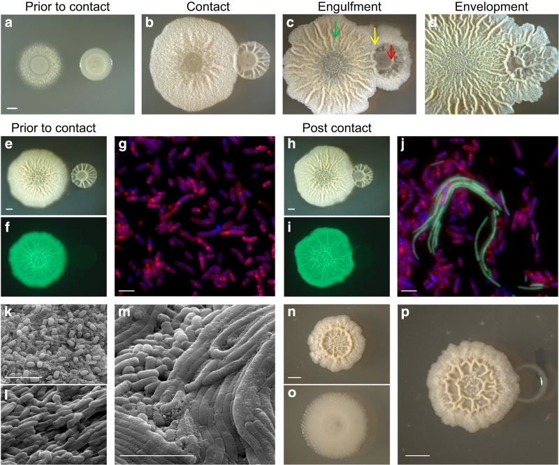
Figure 1.
Antagonistic interaction between B. subtilis biofilms and competing Bacillus colonies. (a–m) Biofilms of B. subtilis and B. simplex grown at 30 °C on MSgg biofilm-inducing medium. (a–d) Biofilms of B. simplex were inoculated next to a B. subtilis biofilm at a distance of 0.8 cm. (a) Before contact (day 1), (b) contact (day 2), (c) engulfment (day 3) and (d) envelopment (day 4). Arrows indicate the different regions of the interaction: B. subtilis area (green), interphase (yellow) and B. simplex area (red), Scale bar represent 2 mm. (e–j) Biofilms of B. simplex were inoculated next to a biofilm of expressing GFP B. subtilis harbouring Phyperspank-gfp at a distance of 1 cm (before contact) and 0.8 cm (post contact), and grown for 2 days. (e,h) Bright field colonies images, scale bar represents 2 mm, (f,i) GFP fluorescence images of the colonies in e and h. (g,j) florescent microscope image: green—B. subtilis strain expressing GFP, red—membrane stain FM4–64, blue—4,6-diamidino-2-phenylindole DNA stain. Scale bar represents 5 μm. (k–m) environmental scanning electron microscopy images of B. subtilis and B. simplex biofilms grown for 3 days. Scale bars represent 5 μm. (k) B. subtilis grown separately. (l) B. simplex grown separately. (m) Interaction area of B. subtilis and B. simplex interacting biofilms in the engulfment stage. (n–p) B. subtilis and B. toyonensis colonies grown for 3 days at 30 °C on B4 biofilm medium. (n) B. subtilis grown separately. (o) B. toyonensis grown separately. (p) Interaction between B. subtilis and B. toyonensis, inoculated 0.3 cm apart. Scale bar represents 2 mm.

