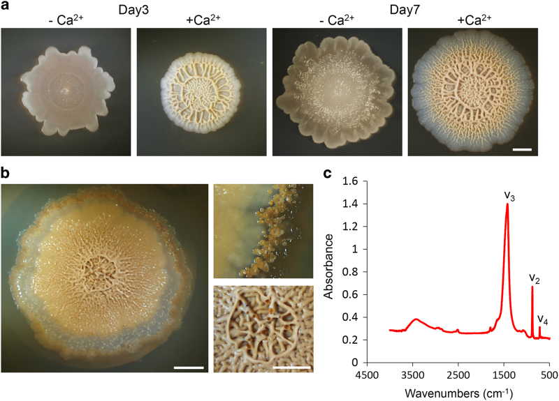
Figure 1.
Complex colony morphology correlates with calcite precipitation in Bacillus subtilis. (a–c) Top view of a colony of an undomesticated strain of wild-type B. subtilis (NCIB 3610). The colonies were grown on solid biomineralization-promoting medium without (a) or with (a and b) a calcium source, for 3 and 7 (a), 21 (b) days, at 30 °C, in a CO2-enriched environment. (b) Top view of a colony (left) and a magnification of calcite crystals at the periphery (right upper) or centre (right lower) of the colony. Images were taken by Stereo microscope with an objective of ×0.5 (a and b, left) or ×1 (b, right) Scale bar corresponds to 2 mm (a and b, left) or 100 μm (b, right). (c) The FTIR spectra of calcium carbonate minerals precipitated at the edges of the colony. ν2 and ν3 indicate characteristic vibrations. The results are of a representative experiment out of five independent repeats.

