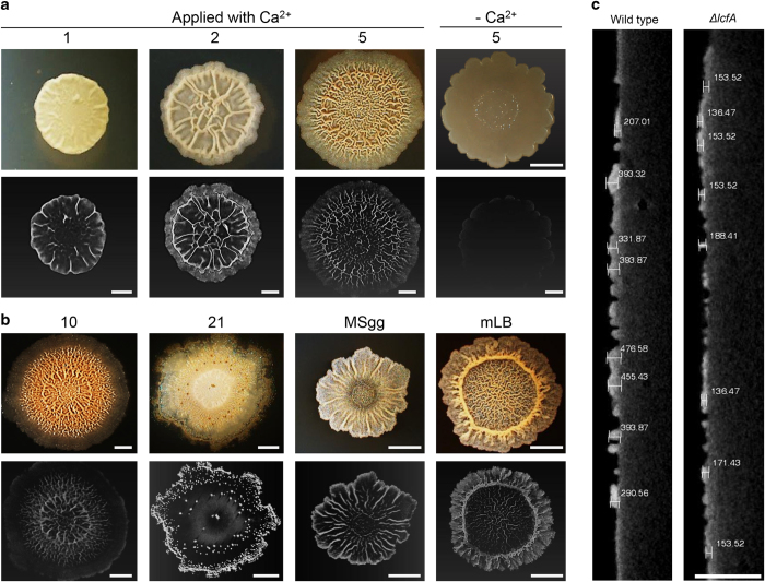
Figure 2.
Calcite and amorphous calcium carbonate have a distinct spatio-temporal organisation within the biofilm. (a, b) Upper panel: Top view of a biofilm of wild-type B. subtilis. The biofilms were grown at 30 °C, in a CO2-enriched environment. (a) On solid biomineralization-promoting medium with a calcium source for 1, 2, 5 days or without a calcium source for 5 days. (b) The biofilms were grown on solid biomineralization-promoting medium with a calcium source for 10, 21 days or on MSgg and mLB medium for 5 days. Images were taken with a stereo microscope with an objective ×1. Scale bar corresponds to 500 μm. Lower panel: MicroCT images of B. subtilis biofilms. Scale bar corresponds to 2 mm. (c) Images representing the thickness of the calcium carbonate buildup underneath the wrinkles of wild-type and lcfA mutant. The images were obtained from the microCT by 2D slice cutting through the biofilm. Scale bar corresponds to 3.5 mm. The results are of a representative experiment out of three independent repeats.

