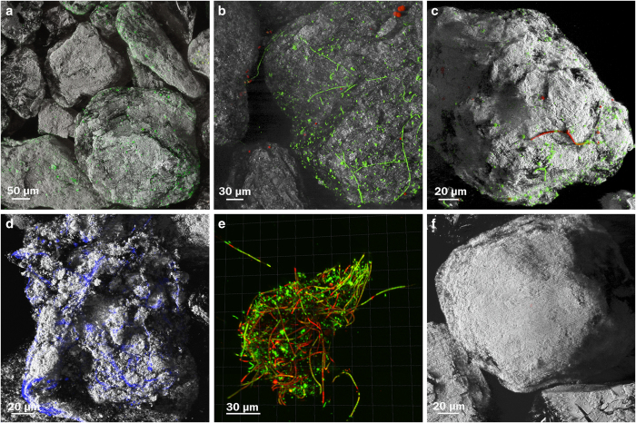
Figure 1.
Representative confocal laser scanning microscopy images of cryoconite sediment with associated microbial communities and biofilm. (a) Green=SYBR Green stained microbes, grey=reflection of the sediment. (b,c) Red=auto-fluorescencing cells, green=SYBR Green stained microbes, grey=reflection of the sediment. (d) Blue=Calcofluor White stained EPS, grey=reflection of the sediment. (e) Red=Propidium Iodide (membrane compromised cells) Green=Syto9 stain (live cells). (f) Control image of combusted cryoconite sediment following described staining protocol, grey=reflection of the sediment.

