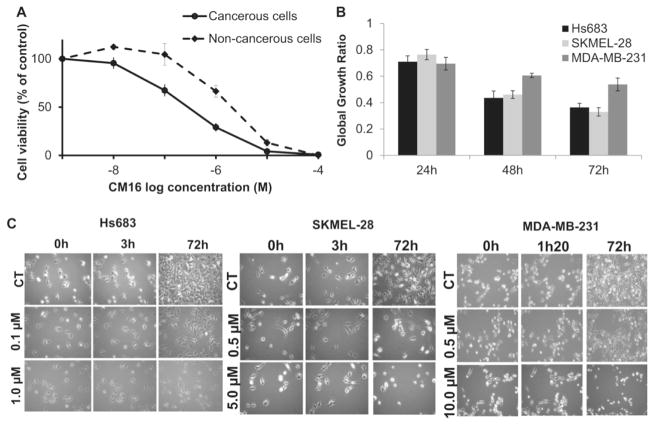
Fig. 3. CM16-induced cytostatic growth inhibition effects in cancer cells.
A: Cell growth inhibition in cancer cells (solid line) versus non-cancerous cells (dashed line) treated with CM16 for 72 h. Cancerous cell lines: Hs683, SKMEL-28 and MDA-MB-231. Non-cancerous cell lines: NHLF and NHDF non-transformed fibroblasts. Data are expressed as the mean of viable cells relative to control (100%)±S.E.M. of the six replicates of one representative experiment. B: Global growth ratio in HS683, SKMEL-28 and MDA-MB-231 cells after 24 h, 48 h and 72 h treatments with CM16 at their GI50. Results are expressed as the mean growth ratio between treated cells relative to control (1)±S.E.M. of three replicates of one representative experiment. C: Videomicroscopy of CM16-induced in vitro effects in Hs683, SKMEL-28 and MDA-MB-231. Figures are representative of one experiment performed in three replicates.

