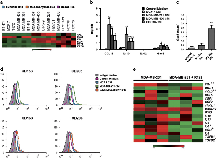
Figure 3.
AXL-overexpressing breast cancer cells promote the polarization of M2 macrophages. (a) Expression analysis of AXL, EMT (CDH1 and VIM), and basal (EGFR, KRT5, and KRT6A) markers in 15 breast cancer cell lines by qRT-PCR. Gene expression levels were visualized in a heatmap. (b) Cytokine levels in the media of macrophages cultured in the absence or presence of MCF-7- or TNBC cell-derived conditioned medium (CM) measured by ELISA. P values were obtained using a two-tailed Student’s t-test (mean±s.d., N=4 experiments; *P<0.05; **P⩽0.01). (c) ELISA analysis of Gas6 in the medium of in vitro polarized human macrophages (Mφ). P values were obtained using a two-tailed Student’s t-test (mean±s.d., N=4 experiments; **P⩽0.01). (d) Flow cytometric analysis of the M2 markers CD163 and CD206 in human monocytes cultured in the absence (pink) or presence of MCF-7-CM (blue), MDA-MB-231-CM (green; upper panel), or R428-treated MDA-MB-231-CM (orange; lower panel) for 6 days. Gray histograms represent staining with isotype controls. The histograms are representatives of five independent experiments. (e) Heatmap showing the effect of R428 on the expression of EMT markers and relevant cytokines/chemokines in MDA-MB-231 cells (three biological replicates were shown). Significant genes were indicated with an asterisks (*P<0.05; **P⩽0.01). ELISA, enzyme-linked immunosorbent assay; EMT, epithelial-to-mesenchymal transition; TNBC, triple-negative breast cancer.

