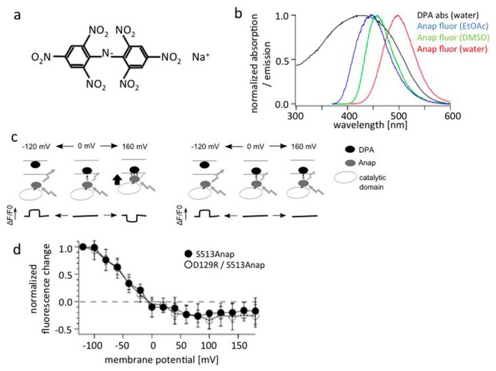
Figure 7.
FRET measurements using DPA and Anap show little change in distance between the membrane and the PD.
(a) Structure of DPA. (b) Absorbance spectrum of DPA (black) and emission spectrum of Anap (other colors). The latter is covered by the former, suggesting DPA serves as a FRET acceptor for Anap emission. (c) The idea behind the use of FRET measurements to detect movement of the Ci-VSP cytoplasmic region toward the membrane. Membrane is labeled with DPA (black ellipse), and the Ci-VSP cytoplasmic region is labeled with Anap (shaded ellipse). DPA is known to translocate between the two membrane leaflets in a voltage-dependent manner. Voltage-dependent changes in the distance between the DPA and Anap are expected to be reported by a decrease in Anap fluorescence upon membrane depolarization (e.g., by a step pulse to 160 mV). (d) FRET measurements with Anap incorporation at S513. This site is known to remain immobile during changes in membrane potential, as judged by the absence of significant changes in the fluorescence of Anap incorporated at the site. The slight decrease of Anap fluorescence most likely reflects voltage-dependent movement of DPA within the membrane. There is no difference between S513Anap and a voltage sensor-defective double mutant (D129R/S513Anap). b, c, d are adapted from [66].

