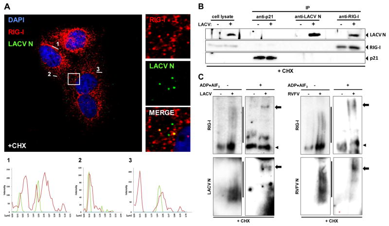
Fig. 4. Interaction of RIG-I with LACV nucleocapsids.
(A) Co-localization analysis. CHX-treated A549 cells were infected with LACVdelNSs and analyzed 5 h later by double immunofluorescence using antisera against LACV N (green channel) or RIG-I (red channel). Cell nuclei were counterstained with DAPI (blue channel). The square area of the inset is digitally magnified on the right hand side. Three fluorescence intensity profiles are shown on the bottom. (B) Co-immunoprecipitation. CHX-treated A549 cells were infected with LACVdelNSs (MOI 10), lysed 5 h later, and subjected to immunoprecipitation (IP) and Western blot analysis using antibodies against p21 (negative control), LACV N, and RIG-I. As input control, 10% of the cell lysate were analyzed in parallel (left lanes). (C) ADP–aluminium fluoride trapping. CHX-treated A549 cells were infected with LACVdelNSs (left panels) or RVFVΔNSs::REN (right panels). At 5 h post-infection lysates were incubated with ADP•AlF3 and analyzed by native PAGE and Western blot using antibodies against RIG-I (upper panels), or viral N (lower panels). Lines indicate oligomers, arrowheads monomers, and arrows point towards high molecular-weight complexes. See also Figs. S11 to S16.

