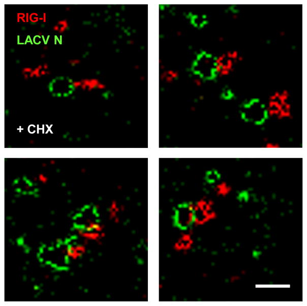
Fig. 7. Super-resolution immunofluorescence microscopy of RIG-I/LACV nucleocapsid complexes.
CHX-treated A549 cells were infected with LACVdelNSs and analyzed 5 h later by GSDIM double immunofluorescence using antisera against LACV N (green channel) or RIG-I (red channel). Four example areas with nucleocapsids are shown. Scale bar 200 nm. See also Fig. S19.

