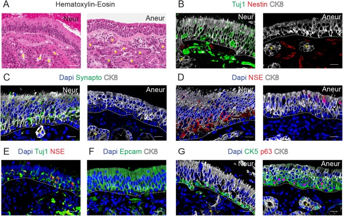
Fig 1. Characterization of the human olfactory epithelium.
Hematoxylin-Eosin staining of the neuronal and aneuronal (Neur, Aneur) samples in A and immunostaining for Tuj1 and Nestin in B, for Synaptophysin (Synapto) in C, for NSE in D, for Tuj1 and NSE in E, for Epcam in F and for CK5 and p63 in G. In most of the panels, CK8 immunostaining was performed and Dapi was used as nuclear counterstaining. Dashed lines indicate basement membrane, yellow asterisks indicate Bowman´s glands and white asterisk indicates axon fibers. Scale bar in A, 100 μm; in B-G, 20 μm.

