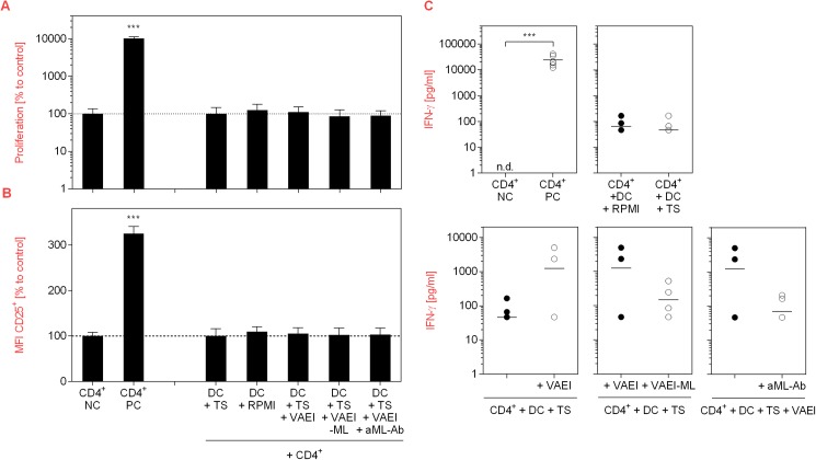
Fig 9. Effects of VAEI-treated DC on T cells after tumor-induced immunosuppression.
Purified human CD4+ T lymphocytes were cultured in the presence of medium (NC) or stimulated with phytohemagglutinin-L (PC; 10 μg/ml). Co-cultivation of T cells was carried out with unstimulated DC treated with 10% RPMI 1640 medium (CD4+ + DC + Medium), 10% tumor supernatant of an RCC line (CD4+ + DC + TS+), TS and VAEI (CD4+ + DC + TS + VAEI; Iscador® Qu Spez; 0.5 μg/ml), TS and ML-depleted VAEI (CD4+ + DC + TS + VAEI-ML; Iscador®; 0.66 μg/ml) or TS, VAEI and anti-ML antibody (CD4+ + DC + TS + VAEI + aML-Ab, CD4+; 2.5 μg/ml). (A) Cell division analysis was done using CFSE staining and flow cytometry. (B) CD25 surface marker expression was analyzed as a second indicator for T cell activation. Data from 6 individual experiments are presented as mean ± SD in relation to untreated T cells (NC) or T cells co-cultured with TS-treated DC (DC + TS). (C) IFN-γ release by T cells was analyzed in the supernatants of 6 independent co-culture experiments using cytokine bead array assay. Asterisks indicate significant differences between the groups (*P < 0.05, **P < 0.01, ***P < 0.001). Data points with values below the detection limit are not pictured.

